


1. Introduction
2. Materials and Methods
Sample collection and isolation
Morphological observations
DNA extraction, PCR, sequencing, and sequence analysis
3. Results and Discussion
Taxonomic account
Morphological characteristics
Molecular characteristics
Comparison with published concepts of Diploneis puella and similar taxa
Molecular comparison and limitations
Significance of the Korean record
1. Introduction
Intertidal tidal flats support diverse microphytobenthic communities in which benthic diatoms are prominent components. Because benthic diatoms are widely used in biodiversity inventories and environmental assessment, reliable species-level identification is important for the interpretation and comparison of ecological datasets (Gottschalk 2014; Méndez-Zambrano et al. 2024). However, species-level identification of small benthic diatoms can be difficult when diagnostic characters are associated with fine valve structures that are not readily resolved by light microscopy.
The genus Diploneis Ehrenberg ex Cleve is a distinctive group of naviculoid diatoms characterized by a raphe system accompanied by paired longitudinal canals. Morphological features such as the longitudinal canals, central nodule, raphe endings, striae, and areolae are important characters for species-level identification (Cleve 1894; Noh and Choi 1992; Droop 1996; Lange-Bertalot and Fuhrmann 2016). In Korea, Diploneis has long been recognized as a component of marine and coastal diatom assemblages. Nevertheless, small Diploneis taxa remain difficult to identify using light microscopy alone because diagnostic characters are often associated with fine external and internal valve structures, including the longitudinal canals, areolar system, and internal openings (Noh and Choi 1992; Lange-Bertalot and Fuhrmann 2016; Kim et al. 2021).
Diploneis puella (Schumann) Cleve was originally described as Navicula puella Schumann and was later transferred to Diploneis by Cleve (Schumann 1867; Cleve 1894). Early records included both inland and coastal localities, such as the Pissa River, Pillau harbor, and the Baltic Sea, indicating that the historical concept of this taxon includes freshwater, brackish, and coastal habitats (Schumann 1867; Cleve 1894). Published concepts of D. puella generally describe a small elliptic valve with radiate striae, but valve dimensions and details of the central and longitudinal structures differ among historical and later accounts (Schumann 1867; Cleve 1894; Lange-Bertalot and Fuhrmann 2016).
The taxonomic concept of D. puella has been applied inconsistently in the literature, and its status remains partly unresolved in contemporary treatments. Lange-Bertalot and Fuhrmann (2016) noted that several historical records attributed to D. puella may represent different taxa and described Diploneis puellafallax Lange-Bertalot et Fuhrmann to distinguish populations previously confused with D. puella. This taxonomic history indicates that modern records of D. puella should be accompanied by detailed morphological documentation, preferably including scanning electron microscopy (SEM), to support reliable taxonomic identification.
In Korea, D. puella has been listed in national species checklists (Lee 1988; NIBR 2025); however, detailed SEM-based documentation and DNA sequence data derived from Korean material have not yet been reported. In addition, publicly available sequence data assigned to D. puella remain insufficient for direct molecular comparison at the species level. In this study, we document strain TA13001, isolated from a Korean tidal flat and provisionally identified as Diploneis cf. puella using light microscopy (LM), SEM, and two molecular markers, 18S rDNA and rbcL. This note provides morphological and molecular documentation of the strain and compares its sequences with available Diploneis reference data.
2. Materials and Methods
Sample collection and isolation
A sediment sample was collected from the Geunso Bay tidal flat on the Taean Peninsula, Chungcheongnam-do, Republic of Korea (36°44′12.06″N, 126°10′47.52″E), on 17 July 2021. At the time of collection, water temperature and salinity of the overlying water were measured in the field using an MP556 Multiprobe System (YSI, Yellow Springs, OH, USA). A single measurement yielded a temperature of 33.1°C and a salinity of 27.6 psu. Surface sediment and overlying water were collected using a sterile plastic container and transported to the laboratory under cool and dark conditions.
A single Diploneis cell was isolated from the sediment sample on 19 July 2021, two days after collection, using the capillary method under an Eclipse Ti-U inverted microscope (Nikon, Tokyo, Japan) and transferred to a cell culture flask (SPL Life Sciences, Pocheon, Republic of Korea) containing F/2 medium supplemented with silicate (Sigma-Aldrich Co., St. Louis, MO, USA) at a salinity of 30 psu. The resulting single-cell-derived clonal strain was designated TA13001 and maintained at 20°C under a 14 h light : 10 h dark cycle with an irradiance of 40 μmol photons m-2 s-1. The culture was confirmed to be unialgal by repeated LM observations showing no other microalgae or diatoms; however, it was not axenic.
The strain was originally established as part of a culture-isolation programme, and the original sediment material was not retained for direct morphological examination. Therefore, comparable valves were not examined directly from the field sample. The first subculture was performed four weeks after single-cell isolation, and material from this first subculture was used for morphological examination. This early-culture material was selected to limit potential morphological changes associated with prolonged cultivation.
Morphological observations
Living cells and cleaned frustules from the first subculture of strain TA13001 were observed using an AX10 light microscope equipped with an Imager A2 digital camera system (Carl Zeiss, Göttingen, Germany). Light microscopy observations were performed using a 100× Plan-Apochromat oil-immersion objective lens. Valve length, valve width, and striae density were measured from 10 cleaned valves using ImageJ software version 1.54s. Terminology for valve morphology follows Ross et al. (1979), Round et al. (1990), Droop (1996), Cox (2004), and Lange-Bertalot and Fuhrmann (2016).
For scanning electron microscopy (SEM) observations, material from the first subculture was fixed in 5% Lugol’s solution, filtered through a polycarbonate membrane filter measuring 25 mm in diameter with a 3 μm pore size (Advantec, Tokyo, Japan), and rinsed three times with sterile distilled water. The membrane was dehydrated through a graded ethanol series from 10% to 100%. Samples were dried using tetramethylsilane (Sigma-Aldrich Co.), mounted on aluminum stubs, and coated with gold using an MC1000 ion sputterer (Hitachi, Tokyo, Japan). Specimens were examined using a high-resolution Sigma 500 VP field-emission scanning electron microscope (Carl Zeiss, Oberkochen, Germany).
DNA extraction, PCR, sequencing, and sequence analysis
Genomic DNA was extracted from material of the same first-subculture clonal strain TA13001 used for morphological observations. Genomic DNA was extracted using the DNeasy PowerSoil Pro Kit (Qiagen Inc., Hilden, Germany) according to the manufacturer’s instructions. The 18S rDNA region was amplified using the primer pair 18S-E (5′-AACCTGGTTGATCCTGCCAGT-3′) and 18S-B (5′-TGATCCTTCTGCAGGTTCACCTAC-3′) (Medlin et al. 1988). The rbcL gene was amplified using DPrbcL1 (5′-AAGGAGGAADHHATGTCT-3′) and DPrbcL7 (5′-AAASHDCCTTGTGTWAGTYTC-3′) (Daugbjerg and Andersen 1997). PCR amplification was performed in a total volume of 20 μL using AccuPower PCR PreMix (K-2012; Bioneer, Daejeon, Republic of Korea). Each reaction contained 1 μL of template DNA, 0.5 μL of each primer (10 pmol μL-1; final concentration, 0.25 μM), and 18 μL of nuclease-free water. The premix contained 1 U of Top DNA polymerase, 250 μM of each dNTP, and 1× reaction buffer containing 1.5 mM MgCl2. For the 18S rDNA region, amplification consisted of an initial denaturation at 94°C for 5 min, followed by 34 cycles of denaturation at 94°C for 45 s, annealing at 55°C for 55 s, and extension at 72°C for 2 min, with a final extension at 72°C for 10 min. For the rbcL gene, amplification consisted of an initial denaturation at 94°C for 3 min, followed by 35 cycles of denaturation at 94°C for 1 min, annealing at 55°C for 1 min, and extension at 72°C for 1.5 min, with a final extension at 72°C for 10 min (An et al. 2017).
PCR products were purified using ExoSAP-IT Express PCR Product Cleanup Reagent (Thermo Fisher Scientific, Waltham, MA, USA). Briefly, 5 μL of each PCR product was mixed with 2 μL of the cleanup reagent, incubated at 37°C for 4 min, and subsequently heated at 80°C for 1 min to inactivate the reagent. Purified PCR products were sequenced bidirectionally by Cosmo Genetech Inc. (Seoul, Republic of Korea). Forward and reverse reads were trimmed, assembled, and checked using Geneious Prime version 2025.2.1 (Biomatters Ltd., Auckland, New Zealand). The resulting sequences were compared with sequences available in GenBank using BLASTn searches performed on 20 June 2026. The sequences were deposited in GenBank under accession numbers PZ575168 for 18S rDNA and PZ573723 for rbcL.
Pairwise alignments were performed using Geneious Prime version 2025.2.1. The closest GenBank matches identified by BLASTn were further compared directly with the TA13001 sequences using pairwise alignments. The aligned length, nucleotide differences, gaps, and ambiguous sites were examined, and the original forward and reverse chromatograms of TA13001 were inspected at differing nucleotide positions. For rbcL, the positions of nucleotide substitutions within codons and their effects on the translated amino acid sequence were also examined.
3. Results and Discussion
Taxonomic account
Diploneis cf. puella (Schumann) Cleve 1894
Basionym: Navicula puella Schumann 1867.
Original description and nomenclature: Navicula puella was described by Schumann (1867: 56, pl. 2, fig. 39) from material collected in the Pissa River near Johannisburg and at the port of Pillau in the Baltic Sea. Cleve (1894: 92) subsequently treated the taxon as Diploneis puella, although he cited Schumann’s name with a question mark, indicating uncertainty regarding the correspondence between the two concepts. Cleve described his material as elliptical, 13–25 μm long and 8–14 μm wide, with a large quadrate central nodule, narrow longitudinal furrows of nearly uniform width, and 12–18 costae in 10 μm.
Original localities and type material: Schumann (1867) cited the Pissa River near Johannisburg and the port of Pillau in the Baltic Sea, former East Prussia. No name-bearing type, lectotype, or repository information associated with the original material could be located in the sources examined; consequently, a single type locality cannot presently be established from designated type material.
Material examined: Republic of Korea, Chungcheongnam-do, Taean Peninsula, Geunso Bay tidal flat (36°44′12.06″N, 126°10′47.52″E), sediment collected on 17 July 2021. Strain TA13001 was established from a single isolated cell. Morphological observations were based on material collected from the first subculture, four weeks after isolation.
Strain: TA13001; single-cell-derived clonal, unialgal, non-axenic culture.
Voucher: Two permanent diatom slides (MABIK DI00044259 and DI00044260), an SEM stub (MABIK DI00044261), cleaned material (MABIK DI00044262; preserved in 100% ethanol), and 5% Lugol-fixed material (MABIK DI00044263) from the first subculture of strain TA13001 were deposited in the National Marine Biodiversity Institute of Korea (MABIK), Seocheon, Republic of Korea.
Description: Valves broadly elliptic to nearly oval with rounded apices, 12.2–17.7 μm long and 5.9–8.6 μm wide, with a length-to-width ratio of 1.9–2.2. Striae robust and slightly radiate, 14–16 in 10 μm. The central nodule is small, quadrate to slightly rectangular, and the longitudinal canals are narrow and linear. Raphe branches are straight to nearly straight throughout most of their course. Externally, the proximal raphe endings are slightly expanded and shortly deflected, whereas the distal endings continue into short, curved terminal fissures. Areolae are loculate and externally occluded by cribra. Internally, the striae open into elongated alveolate chambers separated by robust transapical costae (Figs. 1, 2 and S1).

Fig. 1.
Light micrographs of Diploneis cf. puella strain TA13001 isolated from a Korean tidal flat. Images show valve-view cells displaying broadly elliptic to nearly oval valves, rounded apices, narrow axial areas, and robust radiate striae, and a girdle-view cell at the far right. Scale bar = 10 μm
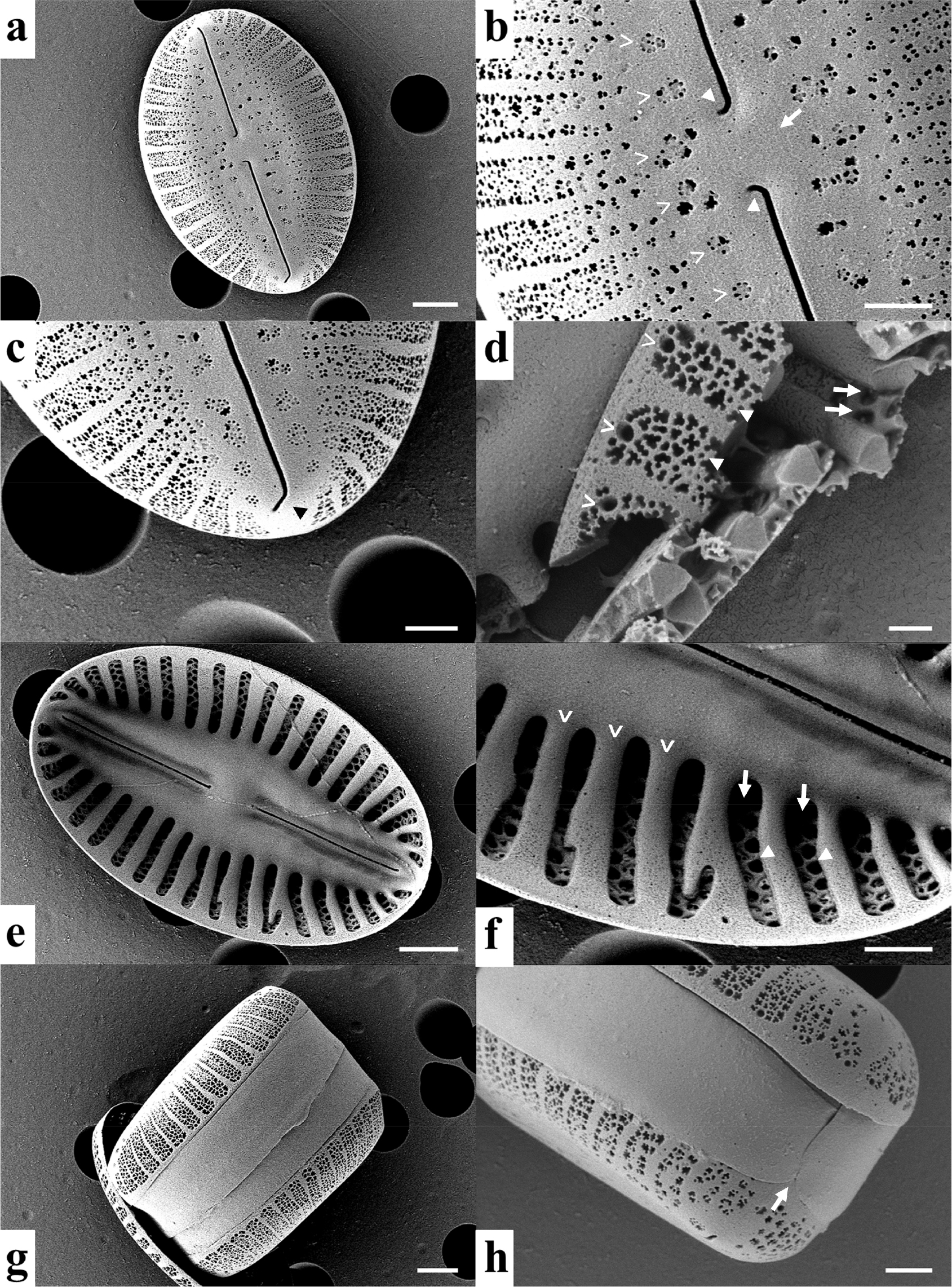
Fig. 2.
Scanning electron micrographs of Diploneis cf. puella strain TA13001 isolated from a Korean tidal flat. a, external valve view. b, central area showing the quadrate central nodule (arrow), external proximal raphe endings (solid arrowheads), and longitudinal canal (open arrowheads). c, valve apex showing the short, curved terminal fissure at the distal raphe ending (arrowhead). d, fractured valve fragment showing loculate areolae (open arrowheads), external cribrate occlusions (solid arrowheads), and pore-like openings near the valve mantle (arrows). e, internal valve view. f, internal striae showing elongated alveolate chambers (arrows), internal areolar foramina (solid arrowheads), and transapical costae (open arrowheads). g, frustule in girdle view. h, valve mantle and girdle region showing a suture-like boundary between adjacent girdle elements (arrow). Scale bars: a, e, g = 2 μm; b, c, f, h = 1 μm; d = 400 nm
Differential diagnosis: Strain TA13001 is morphologically most similar to published concepts of D. puella but is here designated as D. cf. puella because the consistently deflected external proximal raphe endings and short, curved terminal fissures cannot presently be confirmed as part of the intraspecific variation of D. puella. TA13001 is distinguished from D. puellafallax by its narrower valves, broadly elliptic to nearly oval outline, higher stria density, and differences in the central and longitudinal structures. Among other small Diploneis taxa with partially overlapping valve dimensions or morphology, D. rimosa, D. aestuarii, D. modicahassiaca, and D. praetermissa differ from TA13001 in combinations of valve outline and proportions, stria density, central and longitudinal structures, and raphe morphology, as summarized in Table 2.
Habitat and locality: Strain TA13001 was isolated from tidal-flat sediment at Geunso Bay on the western coast of the Republic of Korea. The original sediment sample was collected for culture isolation and was not retained for direct morphological examination.
Previous Korean records of Diploneis puella: The earliest Korean record that could be directly verified in the literature examined here is that of Yi (1983), who explicitly listed D. puella among 208 algal taxa recorded during a survey of the Han River system conducted from April 1979 to June 1981. However, Yi (1983) provided no species-specific illustration, morphometric description, voucher information, or sampling station for D. puella, preventing independent reassessment of that identification. The species was subsequently included in Lee’s (1988) comprehensive checklist of Korean freshwater diatoms, which compiled Korean records published between 1929 and 1986. It has also been retained in later Korean floristic and national checklists. Thus, the name D. puella has a historical basis in Korean floristic literature, but the taxonomic identity of the earlier Korean material cannot be independently verified from the available documentation.
Morphological characteristics
Cells were solitary and motile in culture. Under light microscopy (LM), valves were broadly elliptic to nearly oval, with broadly rounded apices (Fig. 1). Valve length ranged from 12.2 to 17.7 μm, and valve width ranged from 5.9 to 8.6 μm (n = 10). The length-to-width ratio was 1.9–2.2. The axial area was narrow and linear, and the central area was small and slightly expanded around the central nodule. The raphe was filiform and nearly straight. Transapical striae were robust and slightly radiate throughout the valve, becoming more distinctly radiate toward the apices. Striae density was 14–16 in 10 μm. Areolae and internal valve structures were not clearly resolved under LM.
Scanning electron microscopy (SEM) observations showed the external and internal valve ultrastructure in detail (Fig. 2). In external view, the valve was broadly elliptic to nearly oval, with rounded apices (Fig. 2a). The raphe branches were straight to nearly straight throughout most of their course (Figs. 2a and S1a–c). A pair of narrow longitudinal canals was present on both sides of the raphe sternum (Fig. 2b, open arrowheads). The central nodule was small and quadrate to slightly rectangular (Fig. 2b, arrow). Externally, the proximal raphe endings were slightly expanded and shortly deflected near the central nodule (Figs. 2b and S1d). Toward the poles, the distal raphe endings continued into short, curved terminal fissures (Figs. 2c and S1e–g).
The external valve surface showed robust transapical striae, with rounded to irregular pore groups visible under SEM (Fig. 2a–c). A fractured valve fragment showed that the areolae were loculate and externally occluded by fine cribra (Fig. 2d, open and solid arrowheads, respectively). Pore-like openings were also observed near the valve mantle (Fig. 2d, arrows). These observations indicate that the areolar structure is more complex than can be resolved by LM.
In internal view, the valve showed a broad raphe sternum and strongly developed transapical structures (Fig. 2e). The striae opened internally into elongated alveolate chambers (Fig. 2f, arrows). Internal areolar foramina were visible within the chambers (Fig. 2f, solid arrowheads), and adjacent chambers were separated by robust transapical costae (Fig. 2f, open arrowheads). In girdle view, the frustule was rectangular to slightly curved (Fig. 2g). The valve mantle and girdle region were clearly visible, and a suture-like boundary was observed between adjacent girdle elements (Fig. 2h, arrow). Additional SEM observations of separate valves confirmed the straight to nearly straight course of the raphe branches and the repeated configuration of the external proximal and distal raphe endings (Fig. S1). The combination of these morphological characters was further compared with historical and modern descriptions of D. puella and with morphologically similar Diploneis taxa.
Molecular characteristics
A partial 18S rDNA sequence of 1,658 bp and a partial rbcL sequence of 1,428 bp were obtained from the morphologically documented strain TA13001. These sequences were deposited in GenBank under accession numbers PZ575168 for 18S rDNA and PZ573723 for rbcL.
BLASTn analysis of the 18S rDNA sequence showed the highest similarity to Diploneis sp. strain TongYeongLNG circumcp1 (MW327172.1), with 99.76% sequence identity, 100% query coverage, and an E-value of 0.0. Direct pairwise comparison yielded 1,652 identical positions across a 1,656-position local alignment, with one nucleotide substitution, three single-base gap differences, and no ambiguous sites. The three gap-associated differences occurred near the terminal regions of the TA13001 sequence, where chromatogram quality was low, whereas the single internal nucleotide substitution was supported by clear bidirectional chromatogram peaks.
The rbcL sequence showed the highest similarity to Diploneis sp. C isolate TongYeongLNG circumcp1 (MW324596.1) and Diploneis sp. D isolate TongYeongLNG pennE2 (MW324597.1), both with 99.93% sequence identity, 93% query coverage, and an E-value of 0.0. The 93% query coverage reflects that the GenBank reference sequences do not span the full 1,428-bp rbcL sequence obtained for TA13001. Direct comparisons showed that TA13001 differed from each isolate by only one nucleotide substitution across 1,336 aligned positions (1,335/1,336 identical; 99.93%), with no gaps or ambiguous sites. Both differences were supported by clear bidirectional chromatogram peaks and occurred at third codon positions, resulting in synonymous substitutions. The rbcL sequences of circumcp1 and pennE2 differed from each other by two synonymous substitutions across 1,341 aligned positions, also without gaps or ambiguous sites.
Comparison with published concepts of Diploneis puella and similar taxa
The taxonomic concept of Diploneispuella has varied among published accounts, and morphometric information from different sources was therefore evaluated separately rather than combined into a single diagnostic range (Table 1). Schumann (1867) originally described Naviculapuella as a minute elliptical taxon with a valve width approximately two-thirds of the valve length, although the dimensions were reported in historical units. Cleve (1894) subsequently described D. puella as elliptical, 13–25 μm long and 8–14 μm wide, with a large quadrate central nodule, narrow longitudinal furrows of nearly uniform width, and 12–18 costae in 10 μm. Cleve cited Schumann’s taxon with a question mark, indicating some uncertainty regarding the correspondence between the two concepts. More recently, Reavie (2022), synthesizing published accounts of D. puella, reported valve dimensions of 10–27 μm in length and 6–14 μm in width, with 14–18 striae/costae in 10 μm. Strain TA13001 (12.2–17.7 × 5.9–8.6 μm; 14–16 striae in 10 μm) therefore overlaps substantially with these broader published morphometric ranges, although its minimum valve width is slightly below 6 μm. Its length-to-width ratio of 1.9–2.2 nevertheless indicates a relatively elongate valve form compared with the proportions implied by Schumann’s protologue and some later regional accounts. Because morphological observations were based on cultured material, some culture-associated change in valve dimensions cannot be completely excluded. However, the material examined was obtained from the first subculture, four weeks after single-cell isolation, thereby limiting the potential effects of prolonged vegetative cultivation.
Table 1.
Morphological comparison of strain TA13001 with historical and modern published accounts of Diploneis puella
|
Source/ material |
Valve outline |
Length (μm) |
Width (μm) |
L/W ratio |
Striae/ costae |
Central and longitudinal structures |
Raphe / terminal features | Remarks |
| Schumann (1867), original Navicula puella protologue | Elliptical | 2¾–5* |
2/3 of valve length* | ca. 1.5 | 38 in 1/100 line |
Central nodule rounded- elliptical |
Median line faintly marked; terminal nodules scarcely marked |
Original measurements retained separately from later conversions |
| Cleve (1894) | Elliptical | 13–25 | 8–14 | – | 12–18 in 10 μm |
Central nodule large, quadrate; furrows narrow and of equal breadth throughout | Not described |
Alveoli indistinct; attribution to Schumann indicated with a question mark |
| Polaskey (2019), regional account | Nearly oval | 10–12 | 6–7 |
ca. 1.5–1.7 | 14–15 in 10 μm |
Central area small, rectangular; longitudinal canals narrow, linear |
Raphe straight; central ends slightly expanded |
Regional, non-type account |
| Reavie (2022), synthesis of published accounts | – | 10–27 | 6–14 | – | 14–18 in 10 μm | – | – |
Morphometric range summarized from earlier published accounts; not an SEM redescription |
|
TA13001, this study |
Broadly elliptic to nearly oval; apices rounded | 12.2–17.7 | 5.9–8.6 | 1.9–2.2 | 14–16 in 10 μm |
Central nodule small, quadrate to slightly rectangular; longitudinal canals narrow, linear |
Raphe branches straight to nearly straight; external proximal endings slightly expanded and shortly deflected; distal endings continuing into short, curved terminal fissures |
Cultured strain examined by LM and SEM |
*Measurements and stria density from Schumann (1867) are retained in the original historical units.
Among the morphologically similar taxa, Diploneispuellafallax Lange-Bertalot et Fuhrmann requires particular consideration because it was established for material previously confused with D. puella (Lange-Bertalot and Fuhrmann 2016). Its length-to-width ratio (1.7–2.1) overlaps substantially with that of TA13001, and its external raphe shows shortly bent central endings and a gently deflected distal groove. However, D. puellafallax has larger and distinctly broader valves (15–30 × 9–14.5 μm), with no overlap in valve width with TA13001, and a lower stria density of 11–13 in 10 μm. It also differs in its elliptic to more often linear-elliptic valve outline and very small circular to elliptical central area (Table 2). Thus, although valve proportions and some features of the external raphe are similar, the combined differences in valve width, outline, stria density, and central and longitudinal structures distinguish TA13001 from D. puellafallax.
Four additional small Diploneis taxa were considered because their valve dimensions or overall morphology partly overlap with those of TA13001 (Table 2). Among these, the marine species D. rimosa shows particularly close overlap in valve dimensions (12–21 × 6.3–9.7 μm) and has straight external raphe branches with slightly expanded proximal pores. However, it differs from TA13001 in its higher stria density (16–20 in 10 μm), slit-like striae with a broad internal longitudinal rib, hooked terminal fissures, and longitudinal canals that diverge near the central area (Pennesi et al. 2017). D. aestuarii differs in its linear-elliptic valve outline, slight central constriction, deeply depressed area around the central nodule with four areolae, and distinctive configuration of the external raphe endings (Kim et al. 2021). D. modicahassiaca has more strongly elongate, linear-elliptic valves, a higher length-to-width ratio, a higher stria density, and a central area that is not separated from the axial area. D. praetermissa likewise differs in its higher stria density, very small central area, and comparatively long distal raphe endings that curve onto the valve mantle (Lange-Bertalot and Fuhrmann 2016). These combinations of characters distinguish the four taxa from TA13001 despite partial overlap in individual morphometric or morphological features.
Table 2.
Morphological comparison of strain TA13001 with selected morphologically similar Diploneis taxa. A dash indicates that the character was not reported in the cited source
|
Taxon (reference) | Valve outline |
Valve size (μm); L:W |
Striae in 10 μm |
Central and longitudinal structures | Raphe characteristics |
|
TA13001 (D. cf. puella; this study) |
Broadly elliptic to nearly oval, apices rounded |
12.2–17.7 × 5.9–8.6; 1.9–2.2 | 14–16 |
Central nodule small, quadrate to slightly rectangular; longitudinal canals narrow, linear |
Raphe branches straight to nearly straight; external proximal endings slightly expanded and shortly deflected; distal endings continuing into short, curved terminal fissures |
| D. puellafallax (Lange-Bertalot and Fuhrmann 2016) |
Elliptic, more often linear-elliptic; apices broadly rounded |
15–30 × 9–14.5; 1.7–2.1 | 11–13 |
Central area small, elliptic to circular; canal zone narrowly lanceolate |
Raphe filiform, straight; central ends shortly bent and distal ends very weakly deflected unilaterally |
| D. rimosa (Pennesi et al. 2017) | Elliptical |
12–21 × 6.3–9.7; – | 16–20 |
Central area thickened; striae slit-like with a broad internal longitudinal rib; longitudinal canals wide, divergent centrally and convergent apically |
External raphe branches straight; central pores slightly expanded; terminal fissures hooked |
| D. aestuarii (Kim et al. 2021) |
Linear-elliptic, slightly constricted centrally; apices bluntly rounded |
10–21.3 × 5.3–7.3; – | 15–18 |
Central nodule small, square, often indistinct; area around central nodule deeply depressed with four areolae; longitudinal canal narrowly linear |
External central endings bent in opposite directions; terminal endings bent in the same direction |
| D. modicahassiaca (Lange-Bertalot and Fuhrmann 2016) |
Strictly linear-elliptic; apices broadly rounded |
12–16 × 5–6.6; 2.4–2.7 | 16–18 |
Central area not separated; central nodule approximately square or weakly constricted; longitudinal canal zone extremely narrow, linear |
Raphe filiform, straight; central and distal ends inconspicuous in LM; SEM shows shortly bent central ends and slightly deflected distal ends |
| D. praetermissa (Lange-Bertalot and Fuhrmann 2016) |
Elliptic to linear-elliptic; apices broadly rounded |
8–25 × 5.5–7.8; – | 17–19 |
Central area very small, slightly expanded and separated from axial area; axial canals very narrow and slightly arched around central nodule |
Raphe straight; external central ends fish-hook shaped; distal ends long and bent onto the mantle to the same side |
The raphe morphology of TA13001 requires particular caution because detailed SEM observations of type-associated material of D. puella are unavailable. Neither Schumann (1867) nor Cleve (1894) provided a modern ultrastructural description that distinguishes the raphe branches, proximal raphe endings, distal raphe endings, and terminal fissures. A later regional account described the raphe of material identified as D. puella as straight, with slightly expanded central ends (Polaskey 2019), but this material is not type-associated. In TA13001, repeated SEM observations of different valves showed that the raphe branches themselves were straight to nearly straight, whereas the external proximal endings were slightly expanded and shortly deflected, and the distal endings continued into short, curved terminal fissures (Figs. 2b, c, S1d–g). Thus, the observed curvature is restricted to the terminal portions of the external raphe rather than characterizing the course of the raphe branches as a whole.
Because comparable fine-scale SEM observations of type or topotype material are presently unavailable, the raphe configuration observed in TA13001 cannot be demonstrated to fall within the intraspecific variation of D. puella. Although the overall combination of valve dimensions and outline, stria density, the size and shape of the central nodule, longitudinal canals, valve ultrastructure, and raphe morphology is most consistent with the available published concepts of D. puella, the unresolved taxonomic significance of the consistently deflected proximal raphe endings and curved terminal fissures precludes a definitive species-level assignment. We therefore adopt the more cautious designation Diploneis cf. puella for strain TA13001. Future examination of type or topotype material using modern SEM, together with molecular characterization of reliably identified reference material, will be necessary to determine whether the raphe configuration observed in TA13001 represents variation within D. puella or indicates a distinct taxon.
Molecular comparison and limitations
The TongYeongLNG isolates originated from Tong Yeong Station, South Korea, and were included as unidentified Diploneis strains in the molecular dataset of Lobban et al. (2021). However, LM or SEM documentation allowing morphological comparison of the relevant strains with TA13001 was not provided in that study. The very small sequence divergence indicates a close molecular relationship among these Korean Diploneis isolates, but does not by itself establish that they are conspecific or that the TongYeongLNG isolates represent D. puella. Similarly, their provisional identifications as Diploneis sp. C and Diploneis sp. D do not demonstrate that they represent separate species.
The three gap-associated differences observed in the 18S rDNA comparison occurred near low-quality terminal regions of the TA13001 chromatogram and were therefore not interpreted as reliable biological indels. In contrast, the single internal 18S rDNA substitution and the single rbcL substitution in each comparison were supported by clear bidirectional chromatogram peaks. Nevertheless, the extremely small sequence differences among TA13001, circumcp1, and pennE2 cannot be translated into species boundaries in the absence of corresponding morphological documentation for the TongYeongLNG isolates.
At the time of the BLASTn searches, no verified public 18S rDNA or rbcL sequences assigned to D. puella were available for direct species-level comparison. Reliable taxonomic reference libraries require explicit links among molecular sequences, morphological documentation, valid names, and specimen or locality information (Zimmermann et al. 2014). However, barcode reference libraries remain uneven in taxonomic coverage and require expert curation to maintain the reliability of taxonomic assignments, sequence quality, and traceability (Rimet et al. 2019). In Diploneis, recent integrative studies have begun to add molecular data linked to detailed morphology, but molecular sampling remains limited relative to the known and newly recognized morphological diversity of the genus (Jovanovska et al. 2023).
Therefore, the 18S rDNA and rbcL sequences generated here should be interpreted as molecular documentation directly linked to the morphologically characterized strain TA13001, rather than as independent molecular confirmation of species identity. These molecular data are linked to strain TA13001, here designated as D. cf. puella, but do not resolve its relationship to the nominal species D. puella. Such linked morphological and molecular data improve the traceability of Korean Diploneis material and provide a reference for future comparative taxonomic studies.
Significance of the Korean record
Diploneis puella is included in Korean floristic and national checklists, but detailed evidence supporting previous Korean records has remained limited. The earliest Korean occurrence that could be directly verified in the literature examined here was reported by Yi (1983), although the absence of species-specific illustrations, morphometric data, and voucher information prevents independent reassessment of that identification. The present study provides verifiable LM, SEM, 18S rDNA, and rbcL data for strain TA13001, here designated as D. cf. puella because its overall morphology is most consistent with published concepts of D. puella but its raphe architecture cannot presently be reconciled with type- or topotype-associated material.
Accurate documentation of small and morphologically similar benthic diatoms is important for improving the reliability and traceability of biodiversity inventories and environmental datasets. The morphological and molecular documentation provided here therefore establishes a reference for future comparisons of Korean Diploneis material, without implying an ecological role for TA13001 that was not evaluated in this study.
In conclusion, strain TA13001, isolated from a Korean tidal flat, is here designated as Diploneis cf. puella based on its overall morphological similarity to published concepts of D. puella, while recognizing that the taxonomic significance of its consistently deflected proximal raphe endings and curved terminal fissures remains unresolved. Valves of strain TA13001 were characterized by broadly elliptic to nearly oval outlines, rounded apices, narrow longitudinal canals, a small quadrate central nodule, robust radiate striae, loculate areolae externally occluded by cribra, and elongated internal alveolate chambers. Molecular comparisons showed that TA13001 was highly similar to previously deposited unidentified Korean Diploneis strains, although species-level molecular confirmation was not possible because verified public sequence data assigned to D. puella were unavailable. These findings provide traceable morphological and molecular documentation of Korean material assignable to D. cf. puella and establish a reference for future taxonomic reassessment using type- or topotype-associated ultrastructural and molecular data.